


Biomolecules 2023, 13(9), 1338; https://doi.org/10.3390/biom13091338 (registering DOI) - 01 Sep 2023
Abstract
►
Show Figures
SARS-CoV-2, the virus that causes the COVID-19 disease, has been shown to cause immune suppression in certain individuals. This can manifest as a reduced ability of the host’s immune system to effectively control the infection. Studies have reported that patients with COVID-19 can
[...] Read more.
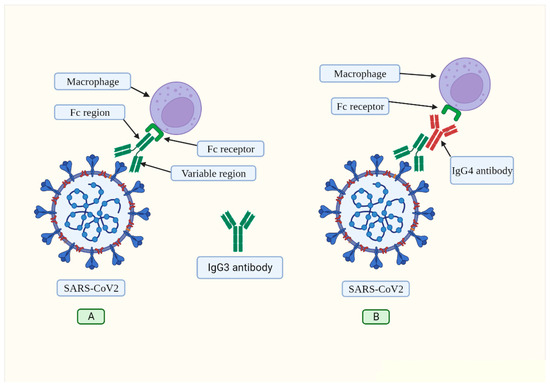
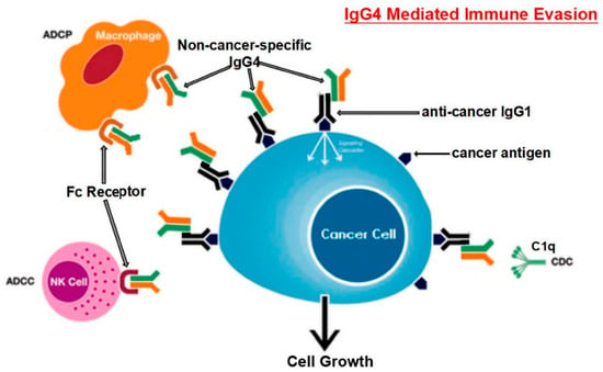
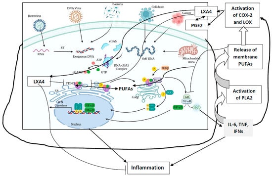
SARS-CoV-2, the virus that causes the COVID-19 disease, has been shown to cause immune suppression in certain individuals. This can manifest as a reduced ability of the host’s immune system to effectively control the infection. Studies have reported that patients with COVID-19 can exhibit a decline in white blood cell counts, including natural killer cells and T cells, which are integral components of the immune system’s response to viral pathogens. These cells play critical roles in the immune response to viral infections, and their depletion can make it harder for the body to mount an effective defense against the virus. Additionally, the virus can also directly infect immune cells, further compromising their ability to function. Some individuals with severe COVID-19 pneumonia may develop a “cytokine storm”, an overactive immune response that may result in tissue damage and organ malfunction. The underlying mechanisms of immune suppression in SARS-CoV-2 are not entirely understood at this time, and research is being conducted to gain a more comprehensive understanding. Research has shown that severe SARS-CoV-2 infection promotes the synthesis of IgG4 antibodies. In this study, we propose the hypothesis that IgG4 antibodies produced by B cells in response to infection by SARS-CoV-2 generate immunological tolerance, which prevents its elimination and leads to persistent and chronic infection. In summary, we believe that this constitutes another immune evasion mechanism that bears striking similarities to that developed by cancer cells to evade immune surveillance.
Full article
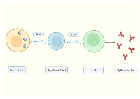
Figure 1


