


by
, , , , , and

Cells 2023, 12(17), 2191; https://doi.org/10.3390/cells12172191 (registering DOI) - 01 Sep 2023
Abstract
Platelets, the smallest cells in human blood, known for their role in primary hemostasis, are also able to interact with pathogens and play a crucial role in the immune response. In severe coronavirus disease 2019 (COVID-19) cases, platelets become overactivated, resulting in the
[...] Read more.
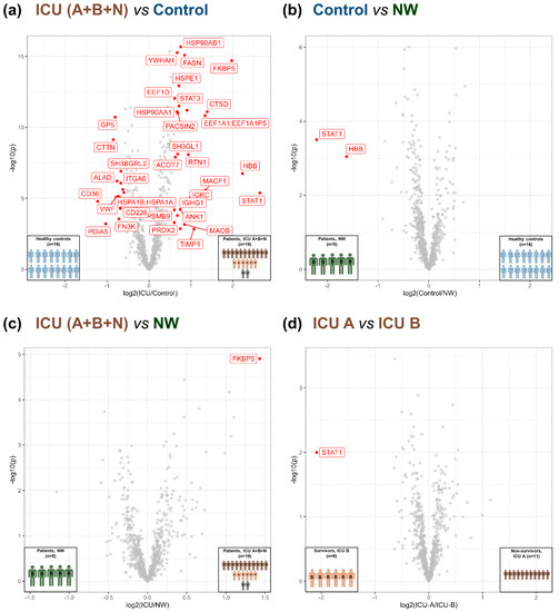
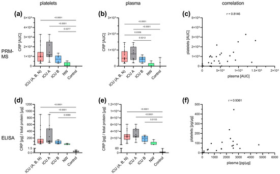
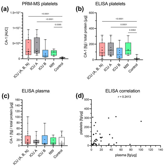
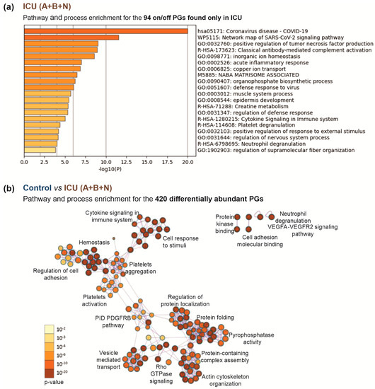
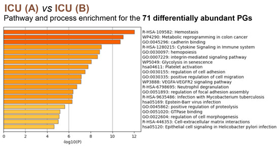
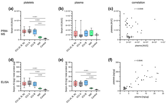
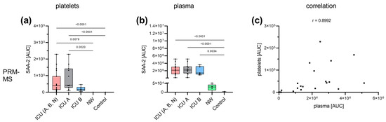
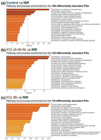
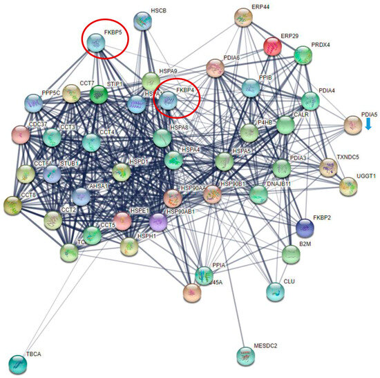
Platelets, the smallest cells in human blood, known for their role in primary hemostasis, are also able to interact with pathogens and play a crucial role in the immune response. In severe coronavirus disease 2019 (COVID-19) cases, platelets become overactivated, resulting in the release of granules, exacerbating inflammation and contributing to the cytokine storm. This study aims to further elucidate the role of platelets in COVID-19 progression and to identify predictive biomarkers for disease outcomes. A comparative proteome analysis of highly purified platelets from critically diseased COVID-19 patients with different outcomes (survivors and non-survivors) and age- and sex-matched controls was performed. Platelets from critically diseased COVID-19 patients exhibited significant changes in the levels of proteins associated with protein folding. In addition, a number of proteins with isomerase activity were found to be more highly abundant in patient samples, apparently exerting an influence on platelet activity via the non-genomic properties of the glucocorticoid receptor (GR) and the nuclear factor κ-light-chain-enhancer of activated B cells (NFκB). Moreover, carbonic anhydrase 1 (CA-1) was found to be a candidate biomarker in platelets, showing a significant increase in COVID-19 patients.
Full article
(This article belongs to the Special Issue Platelet Biology and Functions)
►
Show Figures
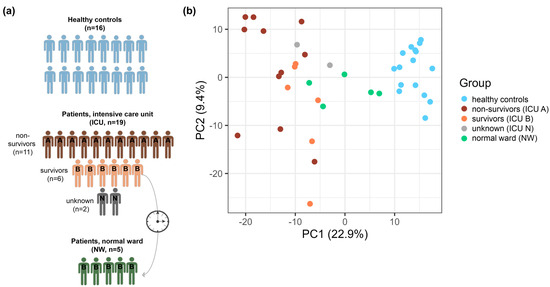
Figure 1


